When the Margin Is Invisible: How 3D Planning Guided a Complex Pelvic Tumor Resection
- Isabelle Têcheur
- Apr 3
- 1 min read


When a tumor invades the ilium, the surgical question is rarely "should we operate?" It is "how much bone do we need to sacrifice?"
Working with Dr. Rémi Garrigue at CHU Reims, we tackled a case of a large iliac tumor where the answer was far from obvious to the naked eye.
The tumor was substantial, but its true extension within the bone is difficult to grasp without a 3D model. Without precise planning, the risk is to unnecessarily widen the resection just to "be safe."
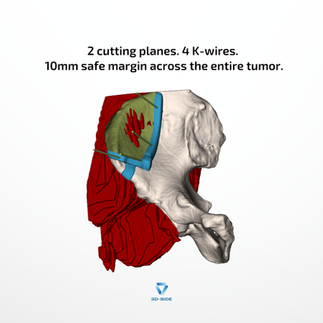
By precisely delineating the tumor on CT and MRI, then modeling its intra-osseous extension in 3D, it became clear that only a small portion of the ilium needed to be resected.
The posterior part of the tumor is particularly critical: without 3D visualization, it remains invisible to the surgeon during the procedure. The saw blade could graze it without anyone knowing.
Planning allows us to see what cannot otherwise be seen, giving the surgeon all the information needed to place the cut exactly where it should be, with a 10mm safe margin guaranteed across the entire tumor.
One patient-specific guide, 2 cutting planes angled for an anterior approach, secured by 4 K-wires. The saw blade knows exactly where to go. No intraoperative guesswork.
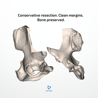
A bone-conserving resection, respected margins, and a surgical procedure prepared down to the last detail before the operating room doors even open.
That is what image-guided, digitally planned surgery looks like.