
A Sacral Tumor Resection: Breaking the Bone Where a Saw Blade Can't Reach
Updated: 2 days ago
Through a posterior approach, the surgeon has good access to the back of the sacrum. But the front side, facing the pelvis, stays hard to reach.
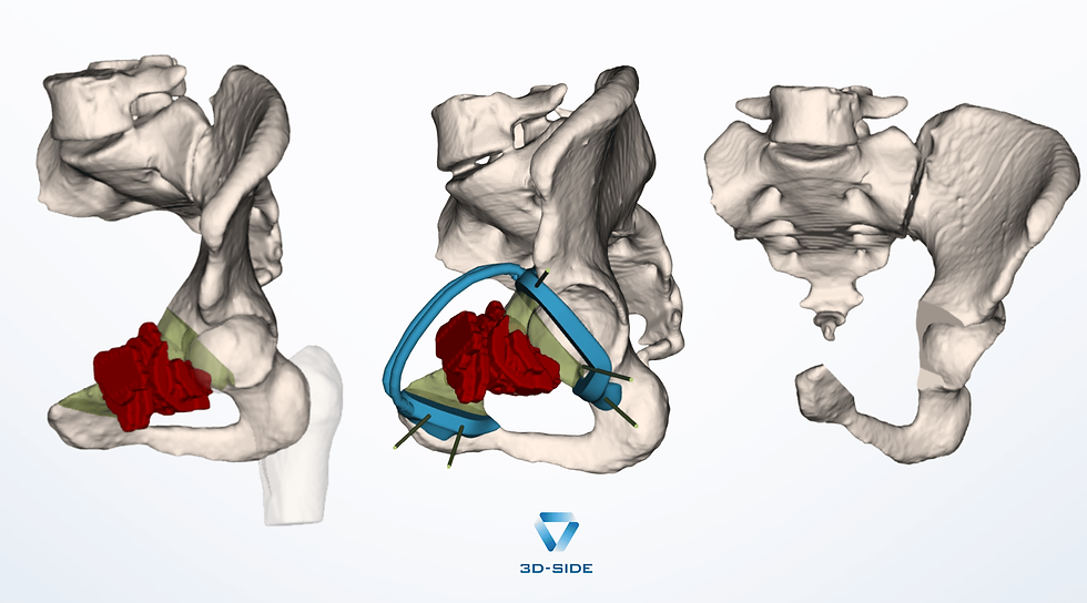
This case involved a sacral tumor resection built on six cutting planes, with a 10 mm safety margin, through a posterior approach. The osteotomy needed to run all the way through the S1 to S4 foramina, close enough to the S1 nerve roots that the plan called for a dedicated step just to protect them. Breaking Bone Where the Saw Can't Follow
A saw can only cut what it can physically touch, so some surfaces are simply out of its reach.
On the endopelvic side of the S1 foramen, the anatomy makes a conventional cut impractical: the surface curves out of reach of a saw blade guided from the posterior approach, and cutting freehand there would come too close to the nerve roots to trust.
Instead of cutting that surface, the guide carries five K-wires aimed specifically at it, driven along the planned resection line to perforate the bone at multiple points.
Once perforated this way, the bone can be broken cleanly along that line rather than sawn through it, keeping the fracture on the exact path the plan defined.
The two most proximal cutting planes run into the same problem and are handled the same way, guided by K-wires rather than cut freehand. Because the osteotomy passes through the S1 to S4 foramina, the distance between the guide and the actual cutting plane changes considerably from one point to the next along the same cut.
For these two planes, that distance is too great for the saw alone, so a chisel completes the cut, following depths marked directly on the guide: 68 mm and 59 mm from the guide's edge to the end of the plane on the endopelvic side. A Guide That Steps Aside for the Laminectomy, and Comes Back
Before the final cuts, the sacral canal needs to be opened at L5 and S1 to keep the nerve roots clear of the resection. Two parallel K-wires, placed early, let the guide be lifted off entirely for the laminectomy and then slid back into exactly the same position afterward, so the plan doesn't need to be re-established once the canal is open.
Closing the Case
The resection fragment is pulled free along a specific distal and posterior path to ease the extraction.
As the cutting planes were planned to spare a maximum of bone and important parts of the sacrum. The planes were only minimally convergent to allow for the specimen extraction. Manual relief cuts were anticipated in case of need.
We thank the surgical team for their trust and for the precision of their feedback throughout the design process.
Facing a similar case? Contact us


Comments